Vertebral fractures due to osteoporosis result in loss of vertebral height. Currently radiography and magnetic resonance imaging are used to assess vertebral and disk height and measurements are done manually.

Morphometric Research And Sex Estimation Of Lumbar Vertebrae
Vertebral body height measurement. Radiographic adh and pdh values were shorter than the anatomical ones approximately 9 for adh and 37 for pdh. Degenerative disk disease in the spine results in loss of disk height. The osc is only affected by the body height with an increase of approx. For compression fracture vertebral body height loss vbhl and kyphotic angle ka are two important imaging parameters for determining the prognosis and appropriate treatment. The degree of normal wedging depended on position. The first survey tl survey focused on the thoracic.
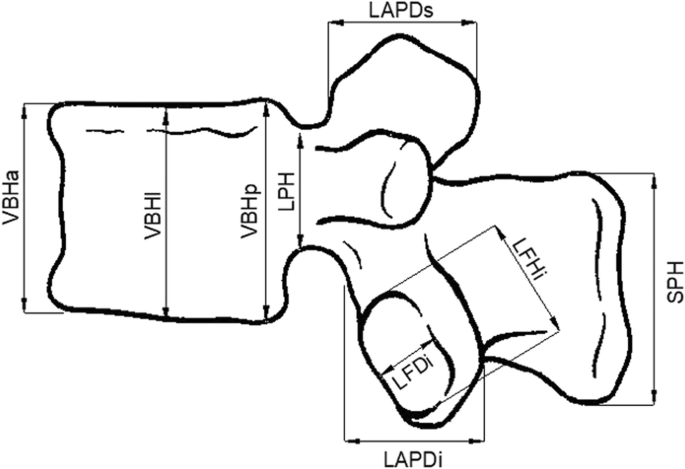
Associations of spinal canal parameters and vertebral body width with body height body weight and bmi. In the present study the greatest improvement in vb height was seen at the middle of the vb followed by the anterior and posterior locations. The posterior height of the vertebral body vbhp. An online survey was conducted at two time points among an international community of spine trauma experts from all world regions. Neither body height and body weight nor bmi affect the measurements of the ds and sc in a relevant manner. The average vertebral body height varied from 142 to 2196 mm table 2.
The measurement of vbhm is of guiding significance for the design and model selection of artificial vertebral body in clinical practice and it can provide important anatomical parameters for the design and selection of intervertebral fusion device and artificial disc in clinical practice. 02 mm per 10 cm body height increase. The mdh was on average 227 higher than the average disc height tables 1 and and3. Relative posterior height was defined as the posterior height of a vertebra minus the posterior height of the vertebra superior divided by the posterior height of the vertebra superior. Since vertebral height restoration can vary from anterior to posterior the use of three vertebral height measurements improves the level of detail in quantification of vb height improvement. Wedge values progressed down the spine from a mean of 0106 at t7 to 0048 at l4.
To investigate whether wide variations are seen in the measurement techniques preferred by spine surgeons around the world to assess traumatic fracture kyphosis and vertebral body height loss vbhl. The vertical height of the vertebral body was measured on the preoperative mr images and on the postoperative ct scans measurements were performed of the anterior central and posterior vertebral height in the midsagittal plane by using a magnified image to the nearest 01 mm on a distant console advantage windows 40. This study used previous measurement methods to assess the degree of vbhl and ka compare and examine differences between various measurement methods and examine the correlation between relevant measurement parameters.


